The Unseen World of Everything
We make sense of the structures, mechanisms and chemical processes shared by all living things. Interested in medical school or a biotech career? We’ll help you get there.
The Science of Life Starts Here
Every living thing has a chemical structure. We research, experiment and pass on the knowledge we find examining all levels of biological organization — including molecular, biochemical, biophysical, structural and organismal.
Our work changes lives. From researching protein interactions related to Alzheimer’s disease to developing vaccines for mosquito-borne viruses, our work impacts human health and well-being around the world.
We’ll help you develop the critical thinking and communication skills needed to succeed in medical or graduate school and in biotech, biomedical or academic careers.
Newly Accredited with ASBMB
RECENT news
Illuminating Science
With support from the new Harold Swaisgood Biochemistry Mentorship Fund, Hali Harwood spent her final undergraduate year at NC State researching a genetic mutation linked to a developmental disorder and two forms of cancer.
Four Incoming CALS Students Named Park Scholars
The four students were selected for the Park Scholarship out of more than 3,400 applicants.
New Software Boosts Accuracy of Tech to Measure Crop Health
A new tool improves the accuracy of electronic devices that measure the color of a plant’s leaves to assess health.
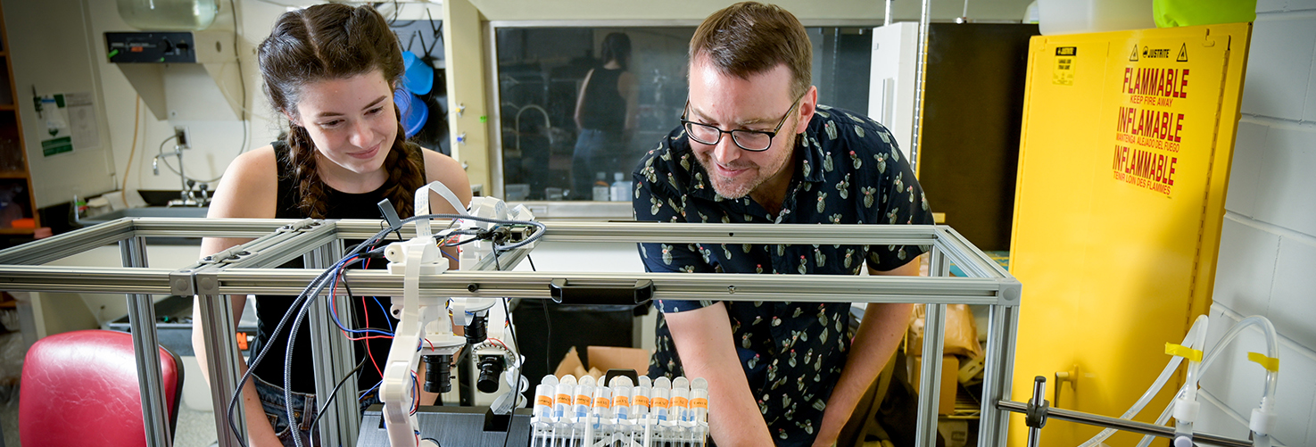
Research Team Awarded UNC ROI Grant to Accelerate Molecular Discovery Using an Autonomous Robotic Lab
An NC State University research team has been awarded funding from the UNC System Research Opportunities Initiative (ROI) to accelerate molecular and biological innovations enabled by next-generation self-driving labs. Self-driving labs…
Domesticating Duckweed, 300 Samples at a Time
What does it take to domesticate a plant? Ryan Sartor is creating unique systems for testing duckweed, an aquatic plant that grows on wastewater.
Simple Trick Could Improve Accuracy of Plant Genetics Research
A simple trick improves the accuracy of techniques that help us understand how external variables – such as temperature – affect gene activity in plants.
Colleen Doherty Wins Fulbright Award to Study Phytomining in England
Doherty will study how plants can be used to mine rare earth elements during a six-month Fulbright Award at the University of York this winter.